The Structure and Function of Fungal Cell Walls: An Overview
Just like plant cells and unlike animal cells, fungi possess a cell wall. The cell wall is nothing but a protective layer within which all of the cellular components are housed. It acts as a protective shield that allows a fungal cell to carry out its functions without threat of destruction from external stresses. While it is easy to think of the cell wall as just a hard shell like that of a tortoise or turtle, this could not be farther from the truth. The cell wall is a highly dynamic and responsive organ of the fungal cell. This dynamic organ is both a target of antifungal drugs and enabler of immune evasion. It also contributes to a majority of the functional benefits we obtained by the consumption of mushroom and mushroom based products. It is constantly changing in response to stimuli and is key to the survival of a fungus. In this blog we will get an idea of the composition of the cell wall.
What is the cell wall
The cell wall of a fungus can be distinguished into two layers: an inner rigid layer and an outer fluid layer. The inner layer forms the structural component of the cell wall. It determines the shape and size of the fungal cell and it prevents the cell from bursting by taking in too much water.
The cell wall is located outside the cell membrane. Ninety percent of the cell wall is composed of polysaccharides. Polysaccharides are long chains of carbohydrate molecules known as monosaccharides bonded together to form a polymer. The most common monosaccharide molecule found in fungal polysaccharides is glucose. Galactose and mannose are also found in lesser proportions. Individual monosaccharides are connected to each other through glycosidic linkages. Polysaccharides are named based on their monosaccharide composition and the nature of the glycosidic bonds between the monosaccharides.
The remaining ten percent of the cell wall may be made up of proteins, lipids and other minor biomolecules. The three major polysaccharide components of the fungal cell wall are: mannoproteins, beta glucans and chitin. Let’s have a look at these three in a bit more detail.
Cell Wall Components

Fig: Cartoon representation of a Candida albicans (yeast) cell wall (Source)
Layer 1: Mannoproteins
Mannoproteins form the outermost layer of the cell wall. This layer consists of polysaccharide chains composed mainly of mannose. A variety of proteins can be found attached to these mannan chains and therefore they are called mannoproteins. This layer is more fluid and porous than the other layers. As the outermost layer, it serves the important function of interacting with the surroundings of the fungal cell. The composition of this layer is highly variable across different fungal species and therefore the least is known about this layer.
Layer 2: Beta Glucans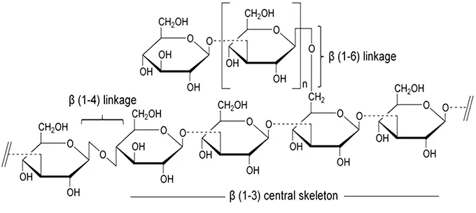
Fig: Representative structure of fungal beta glucans consisting of long 𝛃 (1-3) chains with short branches of 𝛃 (1-6) linked D-Glycopyranosyl units (Source)
The next layer of the cell wall is made up of the famous beta glucans. These form the core structure of the cell wall and form the link between the inner rigid layer and the outer flexible layer. They are composed of long chains of glucose molecules linked to each other via 𝛃 (1→3) glycosidic bonds. These long chains contain short branches also composed of glucose which are linked through 𝛃 (1→6) glycosidic bonds. This results in the formation of a mesh or net-like structure in which other components such as proteins can be embedded.
Layer 3: Chitin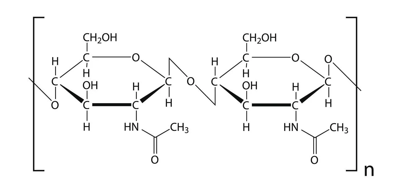
Fig: Representative structure of a chitin molecule consisting of 𝛃 (1-4) linked N-acetyl-Glucosamine (Source)
The innermost layer of the cell wall is composed of chitin. This is the most rigid part of the cell wall. Chitin is the most abundant molecule in the cell wall. Chitin and beta glucans are bound together and form the structural part of the cell wall. The chitin molecules form short chains organised into structures known as fibrils.
These are helical structures that wind around each other (much like a rope). The fibrils of chitin assemble to form tightly packed sheets. Due to this packing it acts as the protective layer of the cell wall, shielding the cell membrane from the harsh outer environment. The beta glucans are found attached to these chitin sheets.
Chitin is the oldest component of the fungal cell wall, surviving through evolution to become the most abundant molecule in the cell wall. Many fungi develop thick chitin layers when the cells are exposed to stress.
The ratios of the different cell wall components are highly variable based on the fungal species. This species to species variation is sometimes used in their identification.
Functions of the cell wall
The cell wall performs many functions in the fungal cell. Its most obvious function is protection. It also acts as the main structural backbone of the fungal cell, determines the shape and size of the cell and helps some fungi evade immune responses.
By forming a multilayered barrier around the fungal cell, the cell wall helps the cell withstand stressful conditions. The cell walls respond to different stresses by altering their composition. For instance, research shows that Aspergillus niger cells can increase the chitin composition of their cell walls by 2.5 times when they are starved. This results in the formation of a thick protective layer that allows the fungus to survive till it encounters a new nutrient source.
As fungi often live in highly variable environments, the composition of molecules such as salts and minerals are constantly changing in their surroundings. The change in salt concentrations lead to the formation of diffusion gradients which result in changing water levels within the cell. In conditions of low salt, the fungal cell swells up due to increased water content. As the cell swells, the cell membrane pushes against the cell wall and exerts pressure on it. These internal pressures can reach as high as 10 MPa in certain fungi. This is 20 times the normal atmospheric pressure! Without the cell walls, the cells would burst.
Fungi which form mycelial threads have elongated cells attached end to end and form highways for the transport of nutrients and water from one end to another. This movement of nutrients is essential to the maintenance of soil ecology.
The fungal cell wall is of immense importance to human health. As it is the interface between the fungus and the external world, it acts as a beacon to our immune system indicating an infection. Most of the major components of the fungal cell wall can be detected by proteins on human immune cells. Upon detection, the immune cells release signaling molecules that inform the immune system of a fungal invasion and trigger an immune response.
To take advantage of this directed immune response, many pathogenic fungi have evolved mechanisms to avoid their detection by altering their cell wall chemistry. These chemical modifications prevent the detection of the fungal cells by our immune cells and allow the fungi to go unnoticed. Other fungi can secrete molecules that act as decoys and prevent the recognition of the fungal cell walls by masking the proteins of the immune system.

Fig: Transmission Electron Microscope images of cell walls from three different fungi showing the variability in cell wall structure (Source)
Synthesis of the cell wall
The formation of the fungal cell wall is a complicated process with multiple steps along the way. Most of the molecules that form the cell wall are assembled within the cell in by the activity of enzymes. These enzymes string together molecules one by one to form the chains that make up the polysaccharides of the cell wall.
Both chitin and glucans are extruded through the plasma membrane of the fungal cell. The basic ingredients for the formation of these molecules are nucleotide diphosphates. In the case of chitin and beta glucans, uridine diphosphate-sugars (UDP) are the carriers that bring together the monosaccharides which form the polysaccharide molecules that are incorporated into the cell wall.
Chitin is synthesised by a family of enzymes known as chitin synthases. These enzymes act on UDP-N-acetylglucosamine. The type of chitin synthase and their relative proportions in the cell determine the structure of the chitin molecules that end up in the cell wall. Similarly, beta glucans are formed the the activity of enzymes belonging to the glucan synthase family. These enzymes string together UDP-glucose molecules.
These enzymes can be found coexisting with each other near the cell wall where they actively extrude these components. Once the polysaccharide molecules are extruded, local enzymes embedded in the cell wall chemically link them to form the complex, multi-layered structure that we observe. The constant change in the number and kinds of proteins in the cell wall affect their structure and help the fungi adapt to various environmental conditions.

Fig: Cartoon representation of fungal cell wall synthesis. Chitin and 𝛃 glucans are synthesised by chitin synthase and 𝛃-1,3-glucan synthase enzymes respectively. The enzymes are embedded in the plasma membrane and extrude polysaccharide molecules to form the fungal cell wall (Source)
Conclusion
Up until the 20th century, the cell wall was assumed to be a passive structural element of cells. However, recent research has highlighted the constantly changing and highly regulated nature of the cell wall. As this structure forms the interface between the external and internal worlds of the cell, it needs to constantly adapt and mediate the survival of the cell. These structures have evolved to be highly complex and can vary immensely between species. However their two core components, chitin and beta glucans, have stood the test of time and are found in all fungi. As such they are responsible for many of the functional benefits observed when we consume fungi. In the next blog we will take a more detailed look at beta glucans and how they help our immune system.
References
Jean-Paul Latgé. 2007 “The cell wall: a carbohydrate armour for the fungal cell” DOI: 10.1111/j.1365-2958.2007.05872.x
E.P. Feofilova. 2010 “The Fungal Cell Wall: Modern Concepts of Its Composition and Biological Function” Microbiology. DOI: 10.1134/S0026261710060019
K. Vega and M. Kalkum. 2012 “Chitin, Chitinase Responses, and Invasive Fungal Infections” International Journal of Microbiology. DOI: 10.1155/2012/920459
Neil A.R. Gow et. al. 2017 “The Fungal Cell Wall: Structure, Biosynthesis, and Function” Microbiology Spectrum. DOI: 10.1128/microbiolspec.FUNK-0035-2016
Ruiz-Herrera and Ortiz-Castellano. 2019 “Cell wall glucans of fungi. A review” The Cell Surface DOI: 10.1016/j.tcsw.2019.100022
De-Oliva Neto et. al. 2015 “Yeasts as Potential Source for Prebiotic β-Glucan: Role in Human Nutrition and Health” DOI: 10.5772/63647
About the author
 Neeladri Chowdhury
As a serial experimentalist and biologist his fascination with fungi began in the Appalachian mountains where he would often marvel at the hundreds of mushrooms seemingly appearing out of nowhere in the fall season. With a master’s in Stem Cell Biology from the University of Minnesota, he has gathered a deep understanding of the processes that govern the regeneration of animal tissues and the development of life. At Nuvedo he aims to unlock the hidden potential of the fungal kingdom.